Imaging renal pH

The kidney is a highly complex organ consisting of welldefined structures that function in a deeply coordinate fashion to allow for fine regulation of pH homeostasis. The role of the kidney in acid-base balance depends on the capacity of the renal tubule to reclaim filtered bicarbonate and to excrete net protons as titratable acids and ammonium.
Although the principal role of the kidney is the maintenance of acid–base balance, current imaging approaches are unable to assess this important parameter and clinical biomarkers are not robust enough in evaluating the severity of kidney damage. Therefore, our lab is developing novel noninvasive imaging approaches to assess the acid–base homeostasis in vivo and to monitor pH evolution following kidney injury. Our lab is also actively involved in the COST Action "PARENCHIMA" CA16103 (Magnetic Resonance Imaging Biomarkers for Chronic Kidney Disease).
We aim to demonstrate the biological validity of renal pH imaging as a novel biomarker of kidney diseases.
Renal pH imaging


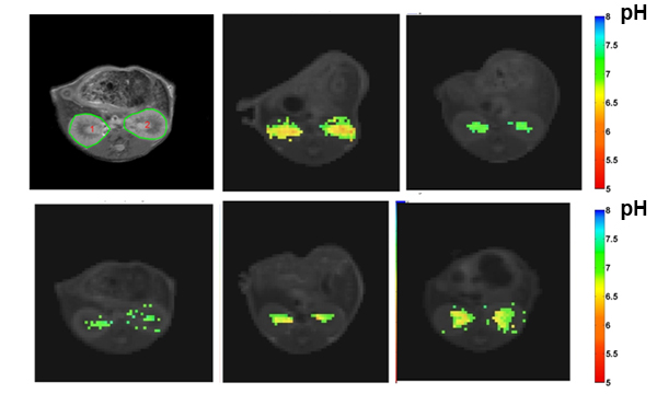
Imaging the pH evolution in an acute kidney injury model


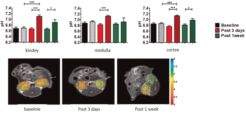
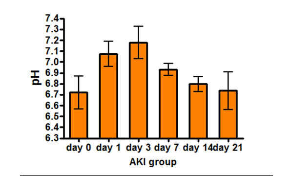
Our previous studies have shown the ability of a iodinated CA, Iopamidol, to measure pH values and induced pH alterations in healthy mice [Longo et al. 2011]. In this study we employed the glycerol-induced AKI model due to the multiple ischemic, toxic, and obstructive tubular insults caused, pathophysiologically similar to the acute tubular necrosis that occurs in humans. According to the elicited damage, this model shows an inherent capability to recover from the damage, therefore allowing to image pH variations both at the acute stage of the injury and during the recovery period. We observed an increase of pH levels reaching a peak at day 3 that correlates well with the corresponding BUN raise and a complete recovery 21 days following the damage [Longo et al. 2013].

We have demonstrated that the use of Iopamidol as a CEST-MRI pH responsive CA provides a good estimate of the kidney pH evolution both in healthy and in pathological conditions. CEST-MRI pH imaging is a suitable method for the noninvasive and longitudinal investigation of renal injury and may provide useful insight on monitoring functional recovery. Moreover, the proposed method provides spatial information on single kidney function and integrity, and not just an average of the kidneys’ functionality as with clinical biomarker such as serum creatinine and BUN.

Single kidney functional pH imaging

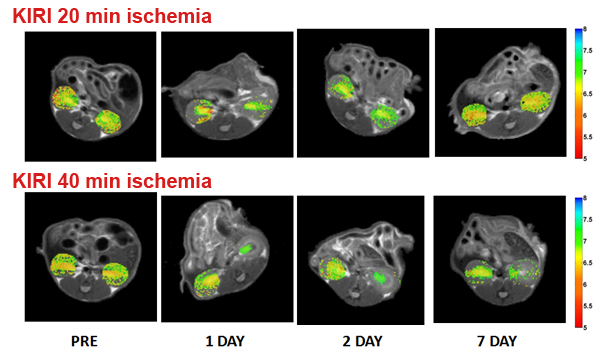
Evaluation of renal pH homeostasis after ischemia reperfusion injury

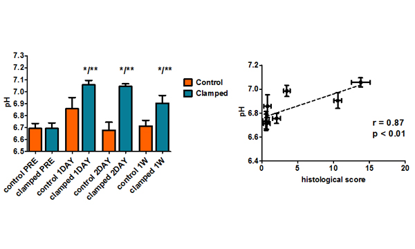
Ischemic renal injury is a severe clinical problem in nephrology and the major cause of acute kidney injury (AKI). We investigated whether MRI-CEST renal pH mapping can detect early renal damage and distinguish AKI recovery from irreversible damage in a unilateral kidney ischemia reperfusion injury (KIRI) model. The advantage of this model is that only one kidney is damaged, whereas the contralateral kidney is not affected. Consequently, in this model of post-ischemic AKI, serum biomarkers are minimally affected, since the contralateral kidney can compensate for the reduced renal functionality. We shown that MRI-CEST pH mapping can detect the early onset of acute kidney damage and distinguish between the recovery and persistence of the damage following unilateral ischemia reperfusion injury in mice. [Longo et al. 2017]. Morover, MRI-CEST renal pH values were significantly correlated with histological scores, reflecting renal damage.

The results reported herein show, for the first time, that MRI-CEST pH mapping allows the noninvasive detection and monitoring of renal function impairment after unilateral ischemia-induced AKI. The derangement of pH regulation was correlated to the decay of renal filtration function and to the severity of the morphological damage.

Dual assessment of kidney perfusion and pH

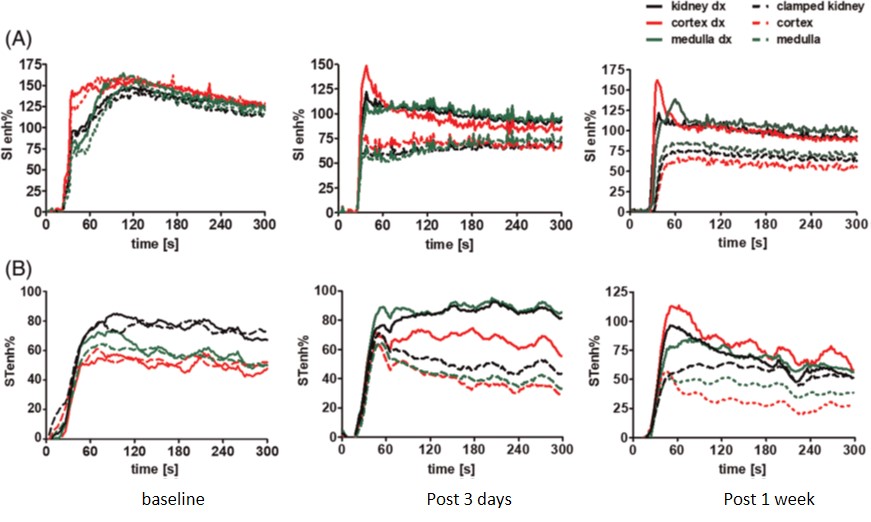
Dynamic CEST-MRI approach in an acute kidney ischemia reperfusion injury murine model

We investigated the exploitation of a single contrast agent injection with a dynamic CEST acquisition for a combined quantification of renal perfusion and pH homeostasis in a unilateral kidney ischemia reperfusion injury (KIRI) model. Thus, a dynamic-CEST approach has been implemented in the acquisition protocol and semi-quantitative estimates (peak, area under the curve AUC and slope) were calculated from the dynamic curves. These measurements have been compared to those obtained with a standard Dynamic Contrast Enhanced (DCE)-MRI approach following Gd-based injection. Compared with the contralateral kidneys, the clamped ones showed a significant decrease in renal perfusion, as measured using the DCE-MRI approach, which is consistent with reduced filtration capability. Dynamic CEST-MRI findings reported similar results, indicating that the clamped kidneys displayed significantly reduced renal filtration that persisted up to 7 days after the damage. [Irrera et al. 2020]. In addition, CEST-MRI pH imaging showed that the clamped kidneys displayed significantly increased pH values, reflecting the disturbance to pH homeostasis.

Our results demonstrate that a single CEST-MRI contrast agent can provide multiple types of information related to renal function and can discern healthy kidneys from pathological ones by combining perfusion measurements with renal pH mapping.