Gd retention in the body

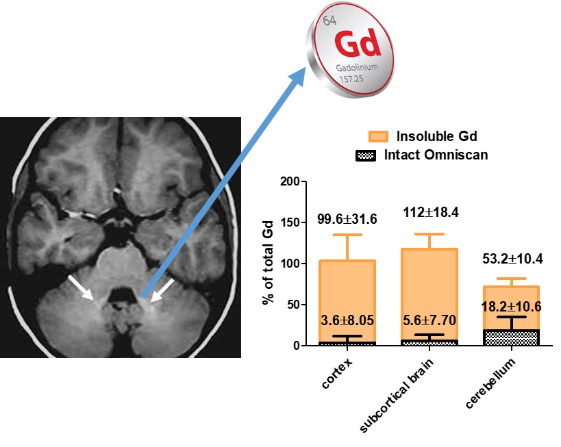
Recently, several studies have shown increased signal intensity on unenhanced T1-weighted MR images in some brain regions in patients with normal renal function who had previously received multiple doses of GBCAs. The observed hyperintense signal has been associated with the retention of small amounts of gadolinium in the brain. There has been evidence that both linear and macrocyclic GBCAs can yield detectable gadolinium traces in the brain, with linear neutral agents leaving greater quantities.

Assessment of the Amounts of Insoluble Gadolinium-containing Species and Intact Gadolinium Complexes after Repeated Administration of Gadolinium-based Contrast Agents

The purpose of this study was to evaluate the speciation of gadolinium containing species in the brain after multiple administrations of gadodiamide and gadoteridol and to quantify the amount of intact gadolinium complexes and insoluble gadolinium-containing species. While the entire fraction of retained gadolinium was in the form of the intact parent complex in animals treated with gadoteridol, the majority of retained gadolinium was in the form of insoluble species and, in part, of a macromolecular system endowed with very high relaxivity in animals treated with gadodiamide [Gianolio et al. 2017].

Insights on the Role of Metal Complex Stability by Comparing Metal Uptake in Murine Tissues Upon the Concomitant Administration of Lanthanum- and Gadolinium-DTPA

The aim of the study was to explore the role of the stability of metal complexes in the processes that lead to the metal retention in the brain and other tissues of mice administered with lanthanides-based contrast agents. This issue was tackled by the simultaneous injection of Gd- DTPA and La-DTPA, which have the same charge and structure but differ in their thermodynamic stability by 3 orders of magnitude [Di Gregorio E et al. 2018].