DCE-MRI for assessing tumor angiogenesis and therapeutic response

It has become evident the key role that tumor vasculature plays in tumor growth and progression. At the same time, a more detailed understanding of the complex parameters that govern the interactions between the tumour and vascular compartments will help to improve anti-angiogenic strategies. Dynamic contrast-enhanced magnetic resonance imaging (DCE-MRI) is the methodology of choice for the evaluation of tumor angiogenesis, following the injection of a Gd-based contrast agent and recording changes in signal intensity along time. This non-ivasive imaging-based approach can provide a detailed view of tumor vasculature, including vessel permeability and plasmatic volume.
Our lab is interested in developing DCE-MRI protocols for accurate assessment of tumor vascuature heterogeneity and in investigating how vasculature changes are correlated with therapeutic response to antiangiogenic treatment.

DCE-MRI for tumor vessel heterogeneity

Imaging therapeutic response to antigiogenic drugs

Dynamic contrast-enhanced magnetic resonance imaging (DCE-MRI) is the methodology of choice for the evaluation of tumor angiogenesis. DCE-MRI allows investigating microvascular structure and function by tracking the pharmacokinetics of an injected Gd-based contrast agent (CA) as it passes through the tumor vasculature. The obtained enhancement patterns reflect vascular perfusion and permeability of the tumor. In the quantitative analysis approach, pharmacokinetic models are applied to contrast agent concentration data to enable estimates of physiological parameters, including plasma volume (vp), forward vascular transfer constant (Ktrans) and the reverse vascular transfer constant (kep). There is an overall agreement in considering tumor heterogeneity as one of the key factors of the disease. Conversely,the values of the DCE-MRI estimates are therefore strongly dependent on how the tumor ROIs are drawn and on the applied statistic analysis. We addressed this limitation by proposing a clustering approach for assessing tumor heterogeneity and thereof changes in the evaluation of the response to a DNA-based antiangiogenic treatment employing a blood-pool contrast agent at 1 T [Longo et al. 2016]. 
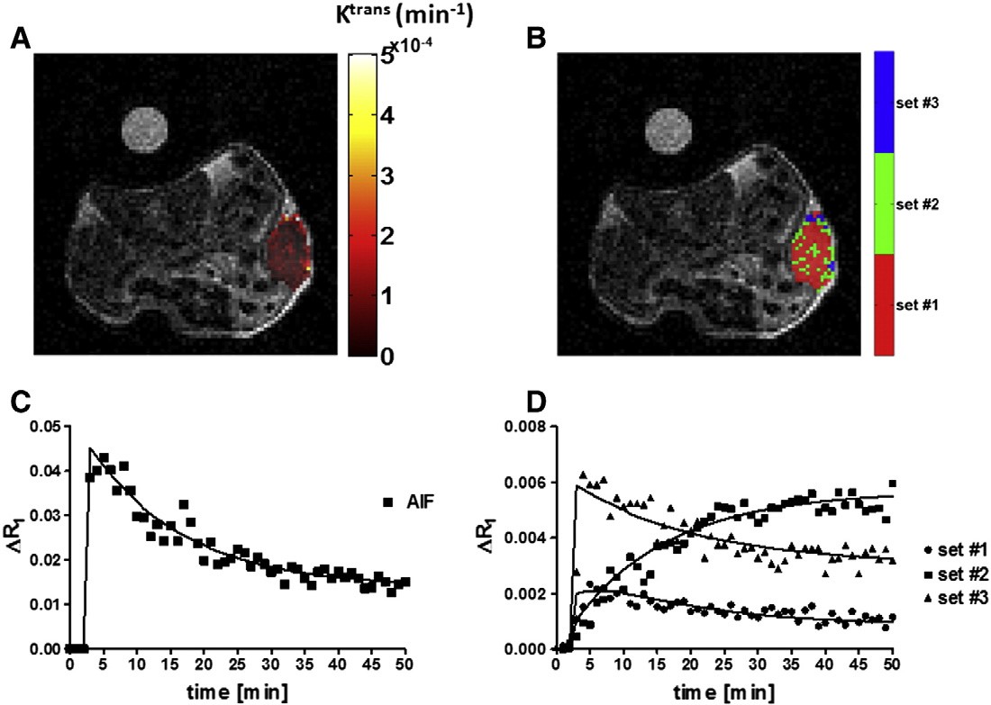
Within the clustering approach, based on a pixel-by-pixel analysis, the whole tumor has been segmented into several sub-regions according to their enhancement/permeability properties. [Longo et al. 2015],

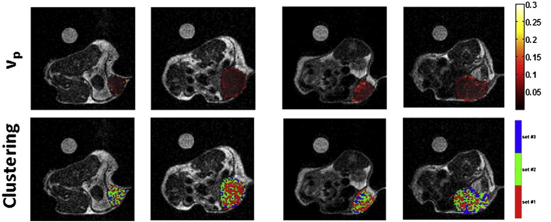
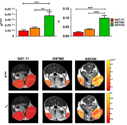
A clustering analysis was performed on a voxel-by-voxel basis to evaluate spatial distribution of tumor Ktrans and vp parametric maps. This study indicates that improvement in the visualization and quantification of heterogeneity in the angiogenic response of tumor to therapy can be assessed by a clustering approach, even though not-homogeneous permeability changes occurred in different areas of a tumor.

We shown that DCE-MRI can also provide assessment of therapeutic response to vaccine targeting tumor vasculature. In fact, a DNA vaccine targeting Angiomotin (Amot, an angiostatin receptors expressed by the endothelia of angiogenic tissues) resulted in tumor growth inhibition that was associated with an increase of vessels diameter and of vessel permeability, as determined by a DCE-MRI approach [Arigoni et al. 2012].

Imaging tumor angiogenesis


Tumor vascularization and angiogenic switch

Tumour progression depends on several sequential events that include the microenvironment remodelling processes and the switch to the angiogenic phenotype, leading to new blood vessels recruitment. Since tumour microvessels exhibit such unique properties, the real time monitoring of alterations in vascularity using DCE-MRI appears to be the most suitable approach to succeed in early breast cancer detection and characterization. We showed that DCE-MRI could detect functional tumour microenvironment changes along the multistep carcinogenesis process observed in the BALB-neuT model, before palpable mass formation, compared to normal mammary glands of BALB/c mice. We demonstrated a significant increase in DCE-MRI biomarkers during malignant transformation. This “angiogenic switch� coincided with an increase in vessel permeability and plasma volume, and histology confirmed that the number of microvessels had increased. These results suggest that DCE-MRI may be a valuable tool for early, non-invasive detection of breast cancer [Consolino et al. 2016]

We exploited a similar approach studying gastrointestinal stromal tumors (GISTs) that develop resistance to the first-line imatinib treatment. Recently, increased vessel density and angiogenic markers were reported in GISTs with a poor prognosis, suggesting that angiogenesis is implicated in GIST tumor progression and resistance. We showed that Imatinib-resistant tumors exhibit increased Ktrans and vp values compared to imatinib-sensitive ones, as confirmed by our ex vivo quantifications of MVD and MDD in GIST430 tumor sections. In addition, a strong positive correlation was observed between MRI and histological estimates [Consolino et al. 2017] .

The current study suggests that the assessment of angiogenesis could be considered a promising new biomarker of response to imatinib treatment.