Investigating tumor pH imaging as a novel cancer biomarker

Tumor cells that live in conditions of hypoxia show an up-regulated glucose metabolism, because of hypoxia-induced shift toward glycolysis. Tumor cells have evolved several sophisticated mechanisms to regulate pH homeostasis: they eliminate acidic catabolites by ion transporters and pumps to preserve a slightly alkaline intracellular pH (pHi), which is optimal for cell proliferation and 
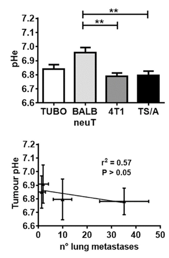
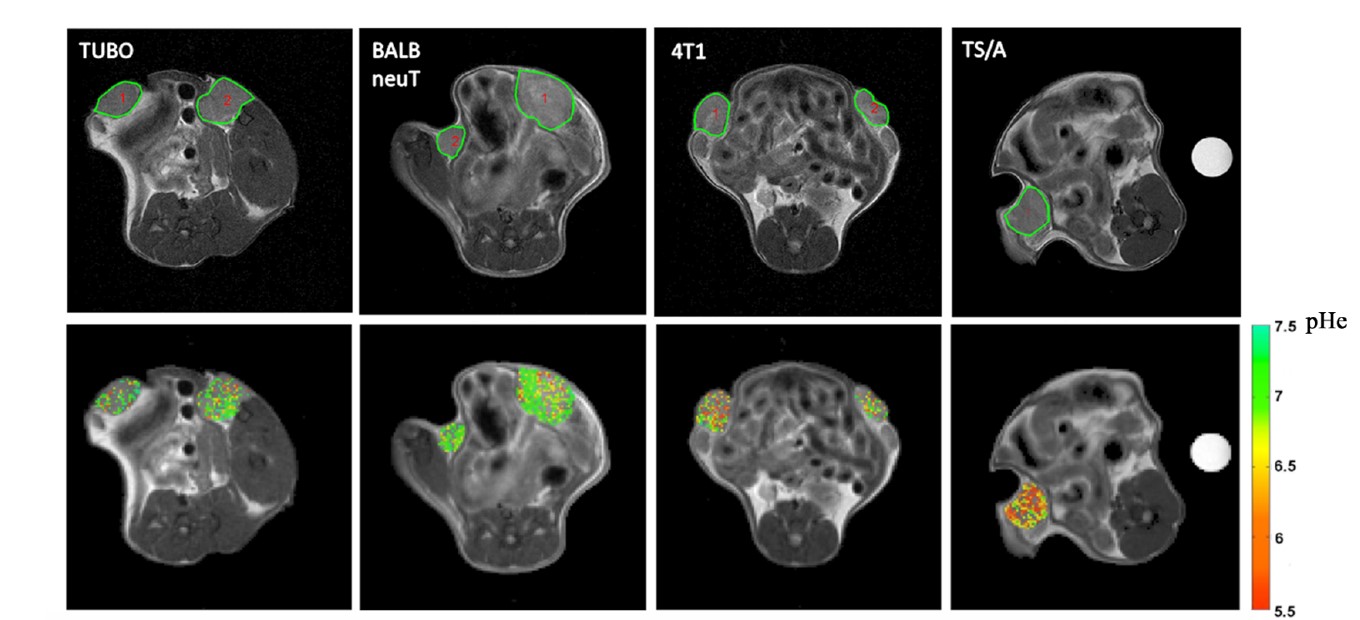
tumor survival.This behavior leads to enhanced acidification of the extracellular pH (pHe) to values in the range 6.5-7.0, which is a distinguishing feature of the tumor microenvironment. Imaging based methods have already been established, at a clinical level, to assess glucose metabolism (by positron emission tomography - PET imaging of 18F-fluorodeoyglucose FDG tumor uptake), providing a formidable tool for evaluating treatment response.
Conversely, despite the excellent studies regarding tumor acidosis, we still do not have an effective acid-base imaging protocol that allows the quantification of extracellular tumor pH and the assessment of pHe related changes following therapeutic treatment.
The focus of our research is to develop non invasive MRI-based tools to assess tumor pHe and to investigate tumor pHe heterogeneity as a potential innovative biomarker for evaluating treatment response to drugs targeting tumor metabolism.

Tumor acidosis


Tumor acidosis and glycolysis


The vast majority of cancers exhibit increased glucose uptake and glycolysis regardless of oxygen availability. This metabolic shift leads to an enhanced production of lactic acid that decreases extracellular pH (pHe), a hallmark of the tumor microenvironment. Despite the fact that aerobic glycolysis and increased extracellular acidification are recognized as hallmarks of solid tumors, no clear evidence of this relationship has been reported so far in vivo. Thanks to the development of accuate CEST pH responsive agents, we were able to study, for the first time, noninvasively the in vivo correlation between tumor 18F-FDG uptake and extracellular pH values in a murine model of HER2+ breast cancer [Longo et al. 2016]. Tumors with higher FDG uptake show lower pHe values, whereas tumors with lower FDG uptake display a less acidic microenvironment

We demonstrated the occurrence of tumor pHe changes that report on acidification of the interstitial fluid caused by an accelerated glycolysis. Combined PET and MRI-CEST images reported complementary spatial information of the altered glucose metabolism. Notably, a significant inverse correlation was found between extracellular tumor pH and 18F-FDG uptake, as a high 18F-FDG uptake corresponds to lower extracellular pH values. These results show how merging the information from 18F-FDG-uptake and extracellular pH measurements can improve characterization of the tumor microenvironment.

pH imaging and tumor therapy

pH imaging for assessing treatment response to cancer metabolism inhibitors

Drugs addressing specific aspects of the deregulated tumour metabolism have been proposed for inhibiting tumour growth and survival. Dichloroacetate (DCA), a mitochondria-targeting small molecule of 150 Da re-establishes the metabolism of pyruvate into the tricarboxylic acid cycle and decreases the lactate accumulation and production. We studied DCA-induced changes in metabolism and tumour acidosis in a murine breast cancer model can be monitored non-invasively by MRI-CEST pH mapping. We observed that lactate production was reduced following DCA treatment both in vitro, just after 24 h, as well as in vivo, with a marked decrease in lactate levels after three days of treatment. Notably, these changes in lactate levels were highly correlated with changes in pHe in vitro and the same strong correlation was found in vivo, likely reflecting the intertwined dependence between glycolysis, lactate levels and tumour acidosis [Anemone et al. 2017].

We demonstrated that MRI-CEST pH imaging is able to detect the early response to DCA by measuring changes in tumour pHe. These results suggest that MRI-CEST pH imaging may serve as a useful imaging biomarker for monitoring changes in metabolism following drugs targeting tumour deregulated glycolysis.

pH imaging and tumor aggressiveness

pH imaging correlates with tumor metastatic potential

It is well known that acidic environment can promote cancer invasion and migration, but few in vivo studies have investigated how tumour pH correlates with cancer invasion. We examined breast cancer cell lines that are distinguished by different metastatic potentials and have been characterized for several markers of aggressiveness and invasiveness. Then, after tumour cell inoculation we studied development of lung metastases and measured in vivo tumour acidosis by MRI chemical exchange saturation transfer (CEST) pH imaging approach [Anemone et al. 2020]. We observed that 4T1 and TS/A primary tumours displayed higher metastatic potential in comparison to the less aggressive TUBO and BALB-neuT ones; this was confirmed by the highest expression of cancer cell stem markers, highlighting their propensity to migrate and invade which coincide with measurement obtained by in vitro assays. [Anemone et al. 2020].

MRI-CEST pH imaging successfully discriminated the more aggressive 4T1 and TS/A tumours that displayed a more acidic pH. Moreover, the observed higher tumour acidity was significantly correlated with an increased number of lung metastases. The findings of this study indicate that the extracellular acidification is associated with the metastatic potential.